

Electron Microscopy
The history of electron optics (ELO) at the Institute of Scientific Instruments was shaped by personalities, whose scientific career had begun during their studies. The students of Prof. A. Bláha – A. Delong, V. Drahoš and L. Zobač – built the first prototype electron microscope in this country, and followed this with the first serially produced instrument (the BS 241). At that time electron microscopes were built in only three countries in the world. In 1954 a functional model of a desktop electron microscope (the Tesla BS 242) was built and it won the Gold Medal at EXPO 1958. Over 1000 of these instruments were produced over a period of 20 years and exported to 20 countries.
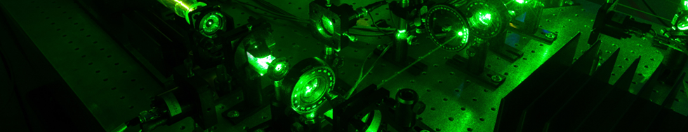
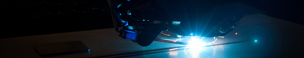
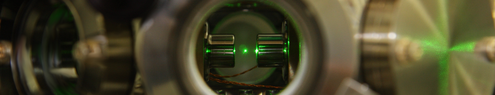
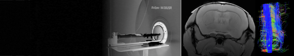
The branch electron microscopy developed significantly in the sixties thanks to the then director Prof. A. Delong and the head of the department of electron optics Prof. V. Drahoš. Unique transmission, emission and scanning electron microscopes were built and, at the same time, the problems of highly stabile current and high voltage sources, problems of vacuum and, subsequently, ultra-high vacuum and analysis of residual gases were resolved. In 1962 the first experiments with electron interferences anywhere in the world were carried out at ISI and were soon applied to various cases. The development and realization of an instrument for electron beam welding was necessary for the construction of ultra-high vacuum devices, and such a device was first realized at the Institute of Scientific Instruments in 1968. The technology of membrane (welded) bellows subsequently proved to be an interesting application. One of the successful transmission microscopes developed in ELO was the TEM TESLA BS 413 microscope with a resolution of up to 0.6 nm and accelerating voltage up to 100 kV, of which 400 were produced by the company TESLA Brno to the end of 1975. At that time non-conventional forms of electron microscopy were also developed, e.g. interference, shadow, Lorentz and tunnel emission microscopy, as well as diffraction under small angles. The first experiments in the world and application possibilities of low-energy electron diffraction were demonstrated with the newly developed ISI electron microscope and were published by A. Delong and V. Drahoš in the journal Nature (1971). The end of the sixties was significant for the achievement of an ultra-high vacuum of 10-6 Pa in the specimen chamber of the emission microscope, which was preceded by the development of a vacuum technique and technology of electron beam welding.
This department also focused on calculations of the magnetic field of lenses and computation of electrons trajectories. Since 1973 the method of finite elements has been exploited for computation of electrostatic and magnetic rotation symmetric lenses. At that time more complex computations were very problematic, as ISI did not own a computer. Computing possibilities did not improve until the nineteen eighties. Regular visits by Professor T. Mulvey from the University of Aston, Birmingham, contributed significantly to the development of this branch and contacts with foreign laboratories.
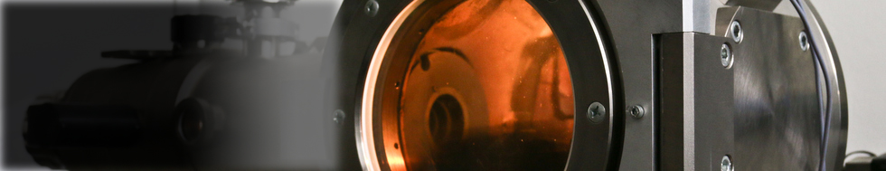
In the middle of the nineteen seventies a team was established, which produced an Auger electron spectrometer in connection with a newly developed scanning electron microscope with a field-emission gun, subsequently produced as the TESLA BS 350. The development of an electron lithograph working with a field-emission gun began at the end of the nineteen seventies. A small series of this instrument was produced by the company TESLA Brno. The device is presently used for research into lithographical techniques in the production of holographic and diffraction structures and for testing preparations for the purposes of microscopy and micro-analysis. The development of new scintillation and cathodoluminescence screens began in the seventies. The introduction of a single crystal of YAG (Yttrium-aluminium-garnet) proved to be a particularly significant success within the scope of these efforts. The detectors based on this principle have become established around the world. In the area of thin layers a world first was achieved by the creation of a multi-layer x-ray mirror with resonance absorption, serving as an analyser during x-ray imaging of biological preparations by phase contrast in the dark field. Unique results in microscopy and diffraction by very slow electrons in scanning electron microscope have recently been presented. Software for electron optics is now improving at an extraordinary rate thanks to the development of information technology. Work in the field of environmental electron microscopy is also proving extremely successful, especially in the area of electron detection in the environment of higher gas pressure.
